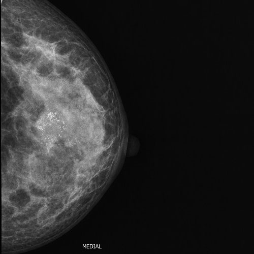
43 year old female with lump in left breast. Mammogram showing mass with scattered microcalcifications within the mass.
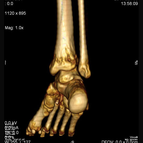
15 year old boy with pain at ankle.
CT with 3D reconstruction shows Osteomyelitis at distal metaphysis of tibia.
CT with 3D reconstruction shows Osteomyelitis at distal metaphysis of tibia.
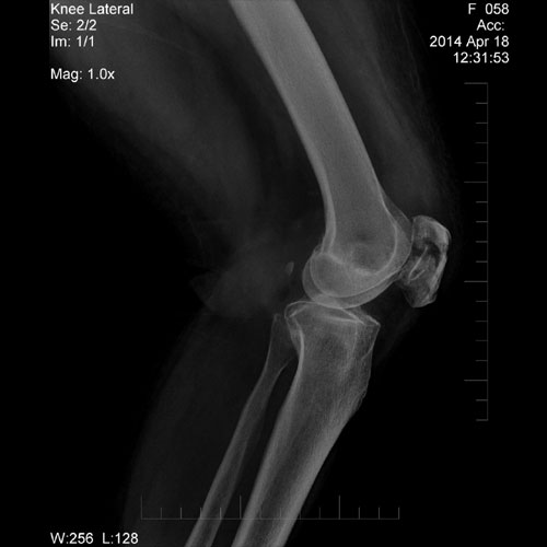
54 year old female with h/o injury; X-Ray of knee joint shows fracture at patella.
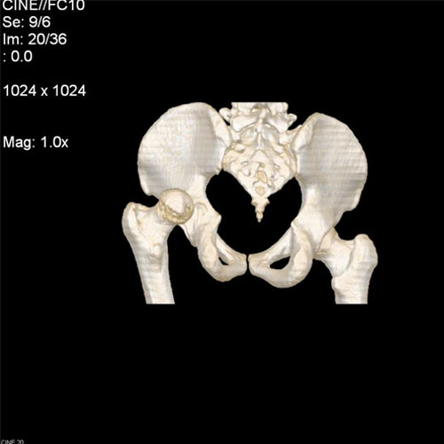
46 year old male, with h/o hip joint pain after epileptic attack. CT with 3D reconstruction of pelvis showing posterior dislocation of hip joint.
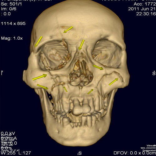
54 year old male with injury to face, CT with 3 D reconstruction shows multiple fractures of facial bones.
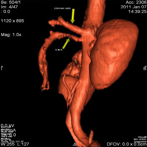
51 year old male with abdomen pain
CT angiogram of abdomen shows partial thrombus at superior mesenteric artery at its origin.
CT angiogram of abdomen shows partial thrombus at superior mesenteric artery at its origin.
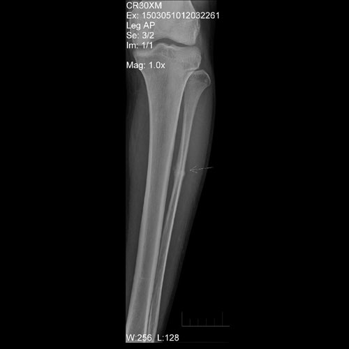
24 year old soldier with pain in mid leg. X Ray of leg shows stress fracture at mid third fibula with callus formation.
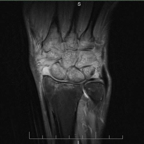
48 year old female with pain in hand. MRI showing Inflammatory arthritis.
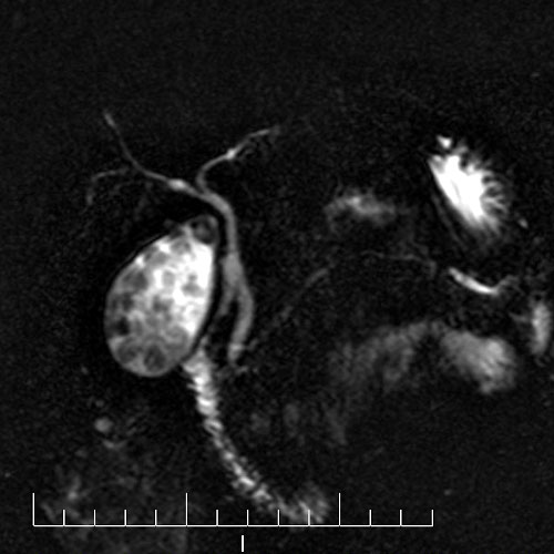
70 year old female with right upper quadrant pain. MRCP showing cholelithiasis
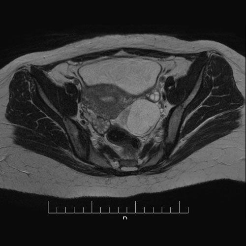
28 year old female with left lower abdomen pain. MRI showing torsion of Fallopian tube.
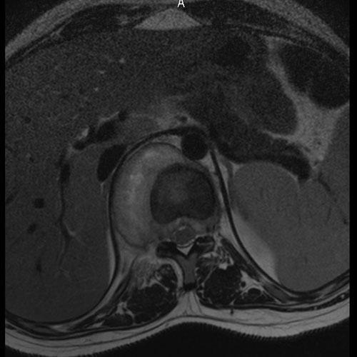
18 year old female with back pain. MRI showing Infectious spondylodiscitis with psoas abscess.
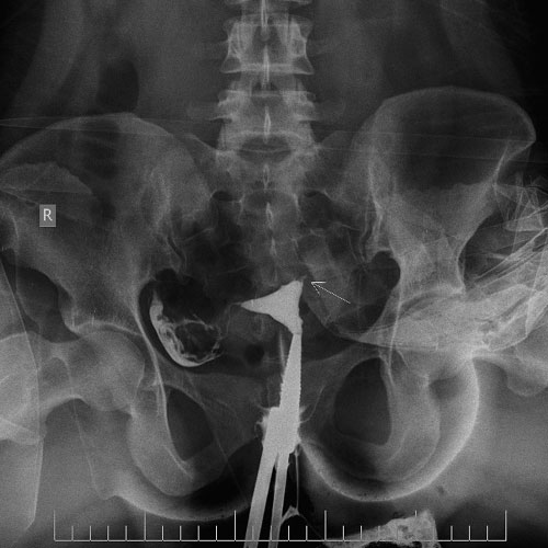
22 year old female with primary infertility. HSG showing blocked tubes on left side.
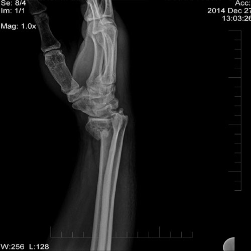
44 year old male with history of fall on outstretched hand. X ray of wrist joint showing Colle’s fracture.
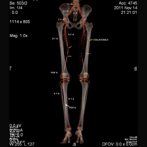
60 year old male with pain in left lower limb.
CT angiogram of both lower limbs shows diffuse atherosclerotic changes. Complete occlusion of proximal superficial femoral artery on left side with reformation of distal third at lower thigh through collaterals.
CT angiogram of both lower limbs shows diffuse atherosclerotic changes. Complete occlusion of proximal superficial femoral artery on left side with reformation of distal third at lower thigh through collaterals.
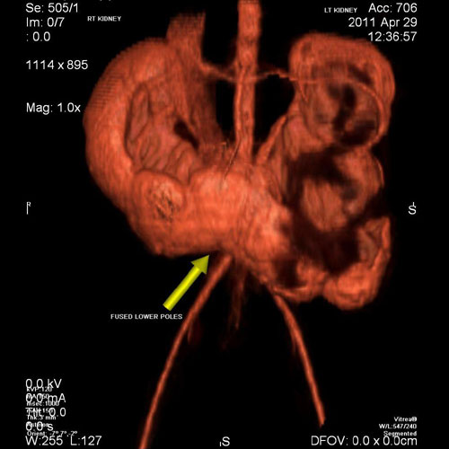
7 year old boy with pain in abdomen on left side. CECT with 3 D reconstruction shows a ‘Horse - Shoe’ shaped kidney with obstructed left side kidney. Horseshoe kidneys are the most common type of renal fusion anomaly. A horseshoe kidney is formed by fusion across the midline of two distinct kidneys. They are connected by an isthmus of either functioning renal parenchyma or fibrous tissue. With a horseshoe kidney, ascent into the abdomen is restricted by the inferior mesenteric artery (IMA) which hooks over the isthmus. Hence horseshoe kidneys are low lying.