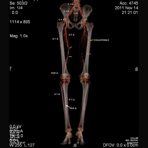
60 year old male with pain in left lower limb.
CT angiogram of both lower limbs shows diffuse atherosclerotic changes. Complete occlusion of proximal superficial femoral artery on left side with reformation of distal third at lower thigh through collaterals.
CT angiogram of both lower limbs shows diffuse atherosclerotic changes. Complete occlusion of proximal superficial femoral artery on left side with reformation of distal third at lower thigh through collaterals.
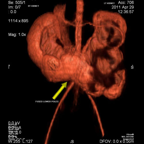
7 year old boy with pain in abdomen on left side. CECT with 3 D reconstruction shows a ‘Horse - Shoe’ shaped kidney with obstructed left side kidney. Horseshoe kidneys are the most common type of renal fusion anomaly. A horseshoe kidney is formed by fusion across the midline of two distinct kidneys. They are connected by an isthmus of either functioning renal parenchyma or fibrous tissue. With a horseshoe kidney, ascent into the abdomen is restricted by the inferior mesenteric artery (IMA) which hooks over the isthmus. Hence horseshoe kidneys are low lying.